
Abstract
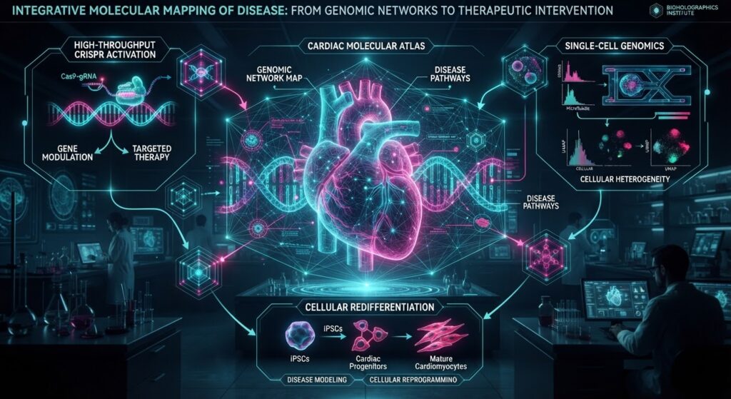
Three recent studies illustrate how high-resolution molecular and cellular mapping is changing the way diseases are investigated. A CRISPRa platform identified genes involved in lymphoma growth and resistance to venetoclax; a CAR-T study showed that temporarily restoring a lost molecular target may increase the effectiveness of immunotherapy against aggressive tumors; and molecular profiling of nearly one million cells provided a more detailed picture of the cellular architecture of hypertrophic cardiomyopathy. None of these studies represents a definitive clinical solution, but together they illustrate a broader transition in modern biomedicine—from studying individual targets toward mapping complex biological systems and the mechanisms that govern their behavior.
Introduction — When Disease Becomes a Map
How can we treat something whose full structure we cannot yet see?
Modern biomedicine is increasingly approaching disease through this question. The study of disease is no longer confined to the level of a single gene or cell. Advances in experimental and molecular technologies now allow researchers to follow the dynamics of thousands of genetic changes and, in some settings, the molecular states of millions of individual cells. This creates increasingly detailed molecular maps of disease1— an important shift because pathological processes are often driven by complex and dynamic mechanisms rather than by a single molecular defect.
A cancer cell may undergo a change that allows it to become resistant to a drug. A tumour may lose the molecule recognised by an immune cell. In heart disease, meanwhile, molecular changes occurring in different cell types may vary depending on the stage and form of the disease.
Three independent studies published in September 2026 address these problems from very different directions. The first uses a new CRISPR2 activation platform to identify genes that contribute to lymphoma growth and drug resistance [1]. The second attempts to temporarily restore a molecular target lost by aggressive thyroid tumours, allowing CAR-T3 cells to recognise them again [2]. The third analyses the molecular profiles of nearly one million individual heart cells to compare different forms and stages of hypertrophic cardiomyopathy [3].
These studies are not extensions of one another, and no direct biological connection has been demonstrated between them. Their broader significance lies elsewhere: the more precisely we can see the molecular and cellular architecture of disease, the more opportunities we have to identify mechanisms that may influence its course.
1. CRISPRa: How Can We Find Genes That Protect Cancer Cells from Drugs?
One of the major challenges in cancer treatment is therapeutic resistance. A drug may initially be effective, yet a subset of tumor cells may find alternative mechanisms that allow them to survive. This raises a fundamental question: which genes give a cancer cell a survival advantage when treatment is applied?
To address this question, researchers from the Olivia Newton-John Cancer Research Institute, WEHI, and Genentech, led by senior author Prof. Marco Herold, used a new CRISPR activation (CRISPRa) platform called Partita.
Partita is a genetic library designed to activate genes individually across the mouse genome, both in cultured cells and in living models. The platform targets each transcription start site with 10 guide RNAs—a high targeting density that contributes to its screening capability. The study was published in Science Advances.
💡 Context: CRISPR Does Not Always Mean “Deleting” a Gene

One of the best-known applications of CRISPR technology is the modification of a specific genetic sequence or the investigation of what happens when a gene loses its function. Modified CRISPR approaches, however, can ask the opposite question:

What happens if a particular gene becomes more active?

This is the basic principle behind CRISPRa—CRISPR activation. Instead of cutting DNA, the system uses a catalytically inactive form of Cas9, known as dCas9, coupled to an activation domain. The complex binds to the promoter region of the target gene and increases its expression without altering the underlying DNA sequence.

This provides a different experimental perspective. A loss-of-function4 approach tells researchers what happens when a gene's function is removed, whereas CRISPRa makes it possible to investigate the consequences of increased or abnormally elevated gene activity.
Irx5 — One Node in Drug Resistance
The researchers performed genome-scale screening in a so-called double-hit lymphoma model against several pro-apoptotic drugs, including venetoclax, nutlin-3a, and etoposide. The most pronounced result was associated with the Irx5 gene: activating Irx5 enabled lymphoma cells to survive in the presence of venetoclax.
To understand the significance of this finding, it is useful to consider how the drug itself works.
Venetoclax is a BCL2 inhibitor. It blocks BCL2, a protein that helps protect cells from apoptosis, or programmed cell death. When BCL2 is inhibited, the cell is pushed toward self-destruction. But BCL2 is not the only survival protein in this family.
The study showed that activation of Irx5 increased levels of BCL-XL (encoded by Bcl2l1), another pro-survival protein from the same family. In other words, when BCL2 was inhibited, the cells appeared to compensate by increasing another component of the cell-survival machinery.
The most direct evidence supporting this interpretation came from experiments in which BCL-XL was inhibited with the compound A-1331852. The same cells then became sensitive to venetoclax again.
The mechanism is thought to involve NF-κB signalling, which is activated downstream of Irx5 and, in turn, increases BCL-XL expression.
A separate screen performed in living mice identified three genes—Runx2, Runx3, and Csf1r—whose activation accelerated the growth of MYC-dependent lymphoma.
💡 Interesting Fact

The same platform therefore addresses two distinct biological questions: which genes accelerate tumour growth, and which genes help a tumour survive treatment?

The distinction matters because the mechanisms driving tumour growth and those responsible for drug resistance are not necessarily the same.

The value of the study therefore lies not only in the identification of individual genes. High-density CRISPRa platforms such as Partita create an opportunity to explore the broader landscape of gene regulation in a systematic way.

There is also an important limitation: the study was performed using mouse models, and the identified genes do not automatically constitute clinical targets. Their relevance in human cells and their potential therapeutic significance require separate validation.
2. When a Tumor Hides: Can a Lost Target Be Restored?
The basic idea behind CAR-T therapy is relatively straightforward: a patient’s T lymphocytes are genetically modified so that they can recognise and destroy cancer cells carrying a particular molecular target.
The approach has transformed the treatment of several blood cancers, but its results in solid tumours have been considerably more limited.
One reason is that a solid tumour can itself lose or reduce the molecular target that CAR-T cells are designed to recognise. In such a situation, the problem may not be that the T cell is insufficiently powerful. The problem may be that its target has disappeared.
Researchers at Mayo Clinic, led by senior authors Saad Kenderian and John Copland III, approached this problem from a different direction. Their study, published in Molecular Cancer, focused on TSHR5 (thyroid-stimulating hormone receptor), a receptor primarily expressed by thyroid cells and therefore an attractive target: its expression in most healthy tissues is limited.
TSHR-directed CAR-T cells have shown activity against differentiated thyroid tumors in which TSHR expression is relatively high. However, anaplastic thyroid cancer—one of the most aggressive forms of thyroid cancer—often has low or nearly absent TSHR expression.
The researchers therefore tried something different. Instead of further modifying the CAR-T cells, they attempted to modify the tumour itself.
Restoring a Lost Target
Loss of TSHR is associated with tumour dedifferentiation, which in turn is linked to excessive activity of the MAPK signalling pathway.
This leads to a straightforward experimental logic: if the pathway is blocked, the tumour cell may partially regain characteristics associated with a more differentiated state. This process is known as redifferentiation6.
The researchers used the MAPK pathway inhibitors trametinib and dabrafenib, which are already approved for the treatment of certain metastatic thyroid cancers.
In patient-derived xenograft7 (PDX) models of anaplastic thyroid cancer, this approach increased TSHR expression and converted tumours from a CAR-resistant state to one that was susceptible to CAR-T cells.
Combining the two approaches produced better tumour control and survival than either treatment alone.
One of the study’s central observations concerns antigen density8: the effectiveness of CAR-T cells may depend not simply on whether a target is present or absent, but also on how much of that target is present on the cell surface. Even a relatively modest increase in target abundance may therefore be sufficient to make a previously resistant tumour susceptible to CAR-T recognition.
Two additional findings are relevant to the practical logic of the approach.
First, MAPK inhibitors did not impair the proliferation or functional activity of the CAR-T cells themselves. This was not necessarily predictable, because MAPK signaling is also active in immune cells.
Second, after treatment with the inhibitors was stopped, TSHR expression began to decline again. Nevertheless, a two-week treatment window was sufficient in the preclinical models to support the combined therapeutic approach.
🔬 GeoCybernetics Perspective

Overcoming resistance to immunotherapy does not always mean making the immune system more aggressively engineered. Sometimes, the problem lies in the target cell itself.

If a tumor cell loses a molecule that an immune cell is capable of recognising, one possible therapeutic strategy is not necessarily to search for a new target, but to temporarily restore the lost one.

This principle remains at the preclinical stage, but conceptually it suggests that therapeutic intervention may be directed not only at the immune system but also at changing the molecular state of the tumour cell itself.

The study is not yet a treatment available for patients. The researchers are continuing the work required to prepare TSHR-directed CAR-T therapy for a first-phase clinical trial.
3. Nearly One Million Cells — A Molecular Map of Hypertrophic Cardiomyopathy
Moving from cancer to a very different biological problem, the third study investigates the molecular architecture of hypertrophic cardiomyopathy (HCM).
In HCM, the heart muscle becomes abnormally thickened and stiff, potentially contributing to heart failure or sudden cardiac arrest. The disease can be associated with pathogenic variants in sarcomere genes, although other cases involve factors that remain incompletely understood.
An international research team from Harvard Medical School, Brigham and Women’s Hospital, and Germany’s Max Delbrück Center for Molecular Medicine used single-nucleus RNA sequencing 9(snRNA-seq).
The study was co-led by Yuri Kim and Eleonora Adami, with Christine and Jonathan Seidman and Norbert Hübner among the senior authors.
The researchers analysed heart tissue from 47 patients, covering disease states from early-stage obstructive HCM with preserved systolic function to terminal-stage disease. They compared these data with tissue from healthy donors and patients with dilated cardiomyopathy.
In total, the analysis included molecular profiles from nearly one million individual heart cells. The study was published in Science Translational Medicine on September 16.
💡 Context: What Does a Single-Cell or Single-Nucleus Approach Mean?

Traditional molecular analyses often treat a tissue sample as a whole. In that case, signals originating from different cell types are mixed together, leaving researchers with an averaged molecular picture.

Single-cell and single-nucleus sequencing take a different approach. They allow researchers to examine gene-expression profiles at the level of individual cells or individual cell nuclei.

This is particularly valuable in complex tissues such as the heart, where cardiomyocytes coexist with many other cell types that may participate in disease processes.
Genetic and Non-Genetic HCM Are Not Molecularly Identical
The researchers identified differences between genetic and non-genetic forms of HCM, as well as between earlier and later stages of the disease.
In genetic HCM, the proportion of cardiomyocytes was relatively lower compared with both healthy tissue and non-genetic HCM. In addition, genes associated with arrhythmia and fibrosis showed increased expression in genetic HCM—two processes that can contribute to disease progression.
The authors suggest that these molecular differences may help explain why arrhythmias and progression toward heart failure are more frequently observed in genetic forms of the disease.
The researchers also identified PRR16 as a gene that may be associated with the cardiomyocyte enlargement characteristic of HCM. Increased expression of PRR16 was supported both by RNA in situ hybridization and in an HCM model based on cardiomyocytes derived from induced pluripotent stem cells.
Another finding concerned differences between disease stages.
In fibroblasts—cells involved in producing and maintaining the extracellular matrix—expression of collagen IV was reduced in early-stage HCM. The authors suggest that this deficiency may contribute to extracellular-matrix destabilization.
This observation is significant because the molecular landscape of HCM does not appear to be confined to cardiomyocytes alone.
When AI Learns to Read the Molecular Map
The final part of the study involved an AI model.
The researchers trained a model on gene-expression data to determine whether different disease states could be distinguished from one another. The model was able to differentiate early from late HCM, HCM from dilated cardiomyopathy, and genetic from non-genetic HCM.
Particularly interesting was the finding that similar differentiation was possible using only fibroblast gene-expression data—cells that are not the primary disease target.
The authors themselves considered this an unexpected result, since cardiomyopathies have traditionally been viewed primarily as diseases of the heart muscle.
This does not mean that a clinically deployable diagnostic AI system has already been developed. More precisely, the study suggests that the cellular microenvironment may contain substantially more information about disease state than can be seen by studying target cells alone.
Three Studies — One Methodological Direction
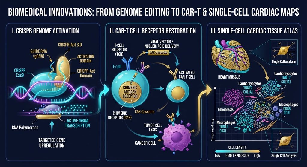
It would be misleading to construct a direct biological connection between these three studies.
Irx5-mediated venetoclax resistance, restoration of TSHR for CAR-T therapy, and the cellular architecture of HCM represent very different biological problems.
Yet they share a deeper methodological similarity:
All three studies attempt to identify important nodes within complex biological systems
In the CRISPRa study, researchers search among thousands of possible genetic nodes for those whose activation can alter cellular behavior.
In the CAR-T study, they ask whether the state of a tumor cell can be changed so that a molecular target that had disappeared becomes visible again.
In HCM, researchers construct a cellular and molecular map capable of distinguishing different forms and stages of disease.
This represents a shift from a single target toward a systems-level map.
🔬  GeoCybernetics Perspective

The creation of a molecular map may sound impressive, but a map and a treatment are not the same thing.

A map shows us where a change occurs.

The next—and considerably more difficult—question is: what causes that change, what is its consequence, and which node can be altered safely and therapeutically?

A resistance-associated gene identified through CRISPRa may become a promising research target, but it was identified in a mouse model and its significance in humans still needs to be established.

Restoring TSHR for CAR-T therapy is an intriguing preclinical strategy, but we do not yet know how it will perform in humans.

The million-cell HCM molecular map provides a more detailed picture of disease architecture, but identifying a new molecular target does not, by itself, mean that an effective treatment against that target exists.

The value of such studies therefore should not be measured only by the individual discoveries they produce. Their significance also lies in the fact that they provide researchers with more detailed working maps of biological systems.
Conclusion — Seeing Disease at Higher Resolution
The three studies published in September 2026 represent three very different biological problems.
The first shows how high-density CRISPRa screening can identify genes involved in tumor growth and resistance to treatment.
The second demonstrates that, in aggressive cancer, it may be useful not only to modify immune cells but also to alter the molecular state of the tumor itself.
The third reveals the complexity of cardiac disease when its cellular architecture is examined across nearly one million individual cellular profiles.
These studies should not yet be interpreted as evidence that new treatments are already close to clinical use. They remain at the preclinical or research stage, and independent validation of the mechanisms, together with evaluation of safety and efficacy in humans, is still required.
But another development may be equally significant.
Modern biomedicine is becoming increasingly capable of studying not only individual molecules, but also the relationships between them, cellular states, and the dynamics of disease.
Perhaps this is the deeper promise of molecular mapping:
the higher the resolution at which we can see disease, the more precisely we can ask where intervention should occur.
The answer to that question remains a subject of research.
But the ability to ask the question itself is already changing.
📚 GeoCybernetics Scientific Glossary & Educational Notes
- Molecular map — A systematic description of changes and relationships involving genes, proteins, cell types, and signalling pathways during disease.
↩︎ - CRISPRa (CRISPR activation)—A CRISPR-based method used to increase the expression of specific genes. It uses a catalytically inactive form of Cas9 (dCas9) coupled to an activation domain, allowing gene expression to be increased without altering the DNA sequence itself.
↩︎ - CAR-T (Chimeric Antigen Receptor T-cell) — An immunotherapeutic approach in which a patient’s T lymphocytes are genetically modified so that they can recognize and destroy cells carrying a specific molecular target.
↩︎ - Loss-of-function — An experimental approach used to study the consequences of reducing or eliminating the function of a specific gene.
↩︎ - TSHR (Thyroid-Stimulating Hormone Receptor)— A receptor protein located on the cell surface that mediates hormonal signaling in thyroid cells.
↩︎ - Redifferentiation—A process in which a tumor cell that has lost differentiated characteristics may partially regain features associated with a more differentiated cellular state.
↩︎ - PDX model (patient-derived xenograft) — A model in which tumor tissue obtained from a patient is transplanted into a laboratory animal. PDX models can preserve some characteristics of the original tumor more faithfully than conventional cell lines.
↩︎ - Antigen density — The amount of a target molecule present on the surface of a cell. In CAR-T therapy, effectiveness may depend not only on whether the target is present, but also on its density.
↩︎ - Single-nucleus RNA sequencing (snRNA-seq)— A method that uses RNA present in individual cell nuclei to characterize gene expression. It is particularly useful for studying the molecular composition of complex tissues.
↩︎
Sources:
- Sarah T. Diepstraten et al. , A high-density CRISPR activation platform for mapping cancer dependencies and resistance pathways ex vivo and in vivo. Science Advances .12, eaec0722 (2026). DOI:10.1126/sciadv.aec0722
- Manriquez-Roman, C., Siegler, E.L., Gleba, J.J. et al. Redifferentiation-enabled TSHRCART cells overcome antigen loss in aggressive thyroid cancers. Molecular Cancer 25, 219 (2026). https://doi.org/10.1186/s12943-026-02744-0
- Eleonora Adami et al.,The molecular landscape of hypertrophic cardiomyopathy across disease stages and genotypes. Science Translational Medicine — 18, eaea2747(2026). DOI:10.1126/scitranslmed.aea2747